

1. Introduction
2. Experimental Procedure
2.1. Scaffold preparation
2.2. Characterization
2.3. Cell studies
3. Results and Discussion
4. Conclusion
1. Introduction
Corneal blindness is one of the leading causes of vision loss worldwide, affecting millions of individuals. Although corneal transplantation is highly successful compared with other organ grafts due to the cornea’s avascular and alymphatic nature, limited donor availability and graft rejection remain major challenges.1) Artificial corneas composed of highly cross-linked collagen have been investigated, but such constructs fail to mimic native corneal biology adequately. Consequently, more effective regenerative strategies are needed. Engineered scaffolds that better replicate the corneal microenvironment have emerged as a promising alternative.2)
Tissue engineering, a branch of biotechnology, aims to develop biological substitutes for repairing or replacing damaged tissues. While early efforts were restricted to two-dimensional cell cultures, recent advances have enabled the generation of complex three-dimensional (3D) tissue constructs. Scaffolds are essential in this context, as they provide a 3D environment that supports cell adhesion, proliferation, and communication. Tissue engineering integrates cells, biomaterials, and bioactive molecules to create functional tissues.3,4)
Corneal tissue engineering approaches that mimic the extracellular matrix (ECM) have been widely studied.5) Both natural and synthetic biomaterials have been evaluated for corneal repair, including collagen,6,7) hyaluronic acid,8) silk fibroin (SF),9) chitosan,10) gelatin,11,12) poly (lactic-co-glycolic acid) (PLGA),13) polycaprolactone (PCL),14) and poly (3-hydroxybutyrate-co-3-hydroxyvalerate) (PHBV).15) Collagen, a key ECM component, exhibits strong bio-adhesion and biocompatibility, making it a widely studied candidate for corneal regeneration.16,17,18) SF has also been extensively investigated. Derived from Bombyx mori cocoons, SF is a natural biopolymer with excellent mechanical properties, biocompatibility, and optical clarity. In addition to its transparency and ease of processing, SF supports corneal epithelial and endothelial cell growth while providing mechanical reinforcement.19,20) Transparent fibroin membranes were fabricated and tested for their ability to support human corneal endothelial cells.
Although coatings with collagen IV, fibronectin-collagen mixture, or chondroitin sulfate improved performance, fibroin substrates still supported lower cell densities compared with standard tissue culture plastic. The vitreous humor (VH) is an avascular, acellular gel primarily composed of glycosaminoglycans (GAGs) such as hyaluronic acid and collagen, along with proteins and vitamins, including albumin, globulins, and ascorbic acid.21,22,23) VH exhibits intrinsic antibacterial properties by inhibiting Gram-positive and Gram-negative bacteria24) and has also been reported to suppress keratocyte differentiation and neovascularization.25) Current research on VH has focused on ophthalmic applications such as intravitreal drug delivery,24) vitrectomy,26) transplantation,22) and diagnostics.27) However, VH hydrogels have poor mechanical strength and degrade rapidly in enzymatic environments, limiting their use in tissue engineering.5)
To overcome these shortcomings, various cross-linkers, including epoxides,28) glutaraldehyde,29) genipin,30) tannic acid,31) and citric acid,32) have been employed. Among them, 1-ethyl-3-(3-dimethylaminopropyl) carbodiimide (EDC) is commonly used to cross-link collagen scaffolds, enhancing both their mechanical strength and biodegradability.16,17)
Taken together, these observations highlight the complementary nature of SF and VH for corneal tissue engineering. It was reported that while SF provides the necessary mechanical strength, structural integrity, and optical clarity, it lacks sufficient biological cues to fully support cellular activity.33,34) Conversely, VH offers a bioactive, ECM-like environment rich in GAGs and functional proteins that can promote cell behavior, but suffers from poor mechanical stability and rapid degradation.34,35,36) Therefore, combining VH with SF represents a rational strategy to integrate the mechanical robustness of SF with the biological functionality of VH.37) This synergistic approach is expected to yield a hydrogel that more closely mimics the native corneal microenvironment while overcoming the individual limitations of each component.
In this study, with a focus on practical application, we aim to develop and evaluate SF-based hydrogels incorporating fish-derived VH as a marine alternative to conventional mammalian sources. Compared to bovine-derived materials, marine-origin VH exhibits a substantially lower risk of zoonotic disease transmission, reduced immunogenicity, and superior immunological compatibility.34,38) Furthermore, it offers significant advantages in terms of sustainability, cost-effectiveness, and large-scale industrial availability.39) Furthermore, differences in the biochemical composition between fish-derived and mammalian VH are anticipated to significantly influence hydrogel network formation and the resulting functional properties, most notably optical transparency, a critical parameter for ocular applications.34,39) Therefore, this work seeks to investigate whether the use of fish VH can provide not only a safer and more sustainable biomaterial platform but also enhanced physicochemical and biological performance compared to previously reported systems.
2. Experimental Procedure
Vitreous gels were isolated from fresh grass carp eyes obtained from a local fish market (Tonekabon, Iran). Eyes were immersed in sterile phosphate-buffered saline (PBS) containing penicillin (500 IU/mL) and streptomycin (500 µg/mL) and processed within 24 h. Connective tissues and anterior segments were removed aseptically, and VH was separated from the lens and ora serrata via a pupil-to-optic nerve incision. Gels were centrifuged at 3,000 × g for 30 min to remove residual cells.26) SF was extracted from raw Bombyx mori silk as described by Chellapandian et al.38) Briefly, SF was prepared from raw Bombyx mori silk fibers by first removing sericin. The raw fibers were boiled in a 0.5 % (w/v) sodium carbonate solution for 30 minutes, then rinsed thoroughly with deionized water to eliminate the sericin coating. The degummed fibers were air-dried and subsequently dissolved in 9.3 M LiBr at 60 °C for 4 h. The resulting solution was dialyzed against deionized water for 72 h using a 12 kDa molecular weight cut-off membrane to remove salts and other small molecules. After centrifugation at 4,000 rpm for 10 min to remove insoluble particles, the regenerated SF solution was stored at 4 °C until use. These conditions ensure consistent removal of sericin and control over the molecular weight of the resulting fibroin. Human corneal epithelial cells (HCECs) were obtained from the National Cell Bank of Iran, Pasteur Institute of Iran, Tehran, Iran.
2.1. Scaffold preparation
Transparent hydrogel solutions were prepared by mixing VH with SF at ratios of 100 % SF, 90/10 SF/VH, and 50/50 SF/VH. Solutions were cast into plastic molds and air-dried at room temperature (~100 µm thickness). Dried membranes were cross-linked in 90:10 ethanol/water containing 1 % (w/v) EDC for 4 h at 4 °C.39) After cross-linking, the membranes were thoroughly washed by immersing them in deionized water for three consecutive cycles of 1 h each, with fresh water replaced at each cycle, to remove any unreacted EDC and urea byproducts. Finally, the washed membranes were air-dried at room temperature before further characterization.
2.2. Characterization
Attenuated total reflectance Fourier transform infrared (FTIR) spectroscopy (ATR-FTIR, Nexus 670, Thermo Nicolet, USA) was used to characterize VH, SF, and SF/VH membranes. Spectra were recorded over the range of 400-4,000 cm-1 at room temperature. To study light transmission and transparency, rectangular membranes (3 × 1 cm, 100 µm thick) were incubated in PBS (1× solution with pH 7.4) at 37 °C for 1 h. Light transmission was measured using a UV-visible spectrophotometer (JENWAY, England) and normalized to PBS baseline. To study swelling and degradation, discs (5 mg, 100 µm thick, and 1 cm diameter) were incubated in PBS (pH 7.4) at 37 °C for 1, 6, 12, 24, and 48 h. After removing the swollen membranes from the buffer, the surface-dried membranes were weighed immediately to determine the sample’s wet weight (W) and compared to dry weight (W0) to calculate equilibrium water content (Wt) according to Eq. (1):
For degradation, discs were incubated in 500 µL PBS containing proteinase K (2 U/mL) at 37 °C. Enzyme solutions were refreshed every other day, and residual mass was expressed as a percentage of initial weight [Eq. (2)].40)
Tensile properties were measured using a uniaxial load machine (SANTAM STM-20, Iran; 20 N load cell, 10 mm/min). Samples (60 × 10 mm, 100 µm) were equilibrated in PBS at 37 °C for 1 h. Maximum tensile strength and elongation at break were calculated from stress-strain curves.
2.3. Cell studies
To study the viability of the membranes, disc membranes (3 mm) were UV-sterilized in 96-well plates and seeded with 5 × 10³ HCECs in 200 µL of Dulbecco’s Modified Eagle Medium (DMEM)/F12 media with 10 % fetal bovine serum (FBS) and 1 % penicillin/streptomycin (100 U/mL). Tissue culture polystyrene (TCPS) served as a control. After 24, 48, and 72 h, 20 µL of 3-(4,5-dimethylthiazol-2-yl)-2,5-diphenyltetrazolium bromide (MTT) solution (5 mg/mL) was added for 4 h at 37 °C. Formazan crystals were dissolved in 100 µL dimethyl sulfoxide (DMSO), and absorbance was measured at 570 nm. Experiments were performed in triplicate. To study cell proliferation, cells cultured for 5 days were fixed with 4 % paraformaldehyde (PFA), permeabilized with 0.1 % Triton X-100, and blocked with 1 % bovine serum albumin (BSA). Nuclei were stained with 1 µg/mL 4′,6-diamidino-2-phenylindole (DAPI) and imaged using fluorescence microscopy (Nikon DXm1200F). Negative controls without primary antibodies were included. Experiments were conducted in triplicate. To study cell adhesion, HCECs (~104 cells) were seeded on 1-cm discs and cultured in DMEM/F12 with 10 % FBS, 1 % penicillin/streptomycin at 37 °C, 5 % CO2. Media were refreshed every 2 days. Samples were fixed in 2.5 % glutaraldehyde, rinsed with PBS, dehydrated in graded ethanol (30-100 %), sputter-coated with gold, and imaged using scanning electron microscopy (SEM; Philips XL3, 25 kV). Data are presented as mean ± SD. Student’s t-test was used for within-group comparisons, and two-way ANOVA with Tukey’s post-hoc test was used for between-group comparisons. Significance was set at p < 0.05 (within-group) and p < 0.01 (between-group).
3. Results and Discussion
In this work, we investigate the use of fish-derived VH as a marine-based alternative to traditional mammalian sources in the development of SF hydrogels. Compared to bovine-derived materials, marine sources are generally associated with lower risks of disease transmission and improved biological acceptance, while also being more sustainable, cost-effective, and readily available for large-scale use. At the same time, differences in biochemical composition between fish and mammalian VH may influence the cross-linking process and the resulting hydrogel structure, which can ultimately affect key properties such as transparency and overall performance. Based on this rationale, the following results examine whether this marine-derived system can offer a safer, more practical, and functionally improved alternative to previously reported hydrogels. In the SF-VH system, EDC-mediated cross-linking is expected to primarily occur between the carboxyl groups of aspartic and glutamic acid residues in SF and the free amino groups present in collagen and other protein components of VH. Additionally, GAGs such as hyaluronic acid and chondroitin sulfate in VH contain carboxyl groups that can potentially participate in cross-linking with amino groups from SF or VH proteins such as collagen and albumin, contributing to the formation of a three-dimensional network [Fig. 1(a)]. Thus, the resulting hydrogel network is stabilized through multiple covalent linkages between SF and VH components, integrating the mechanical robustness of SF with the bioactive, ECM-like nature of VH.
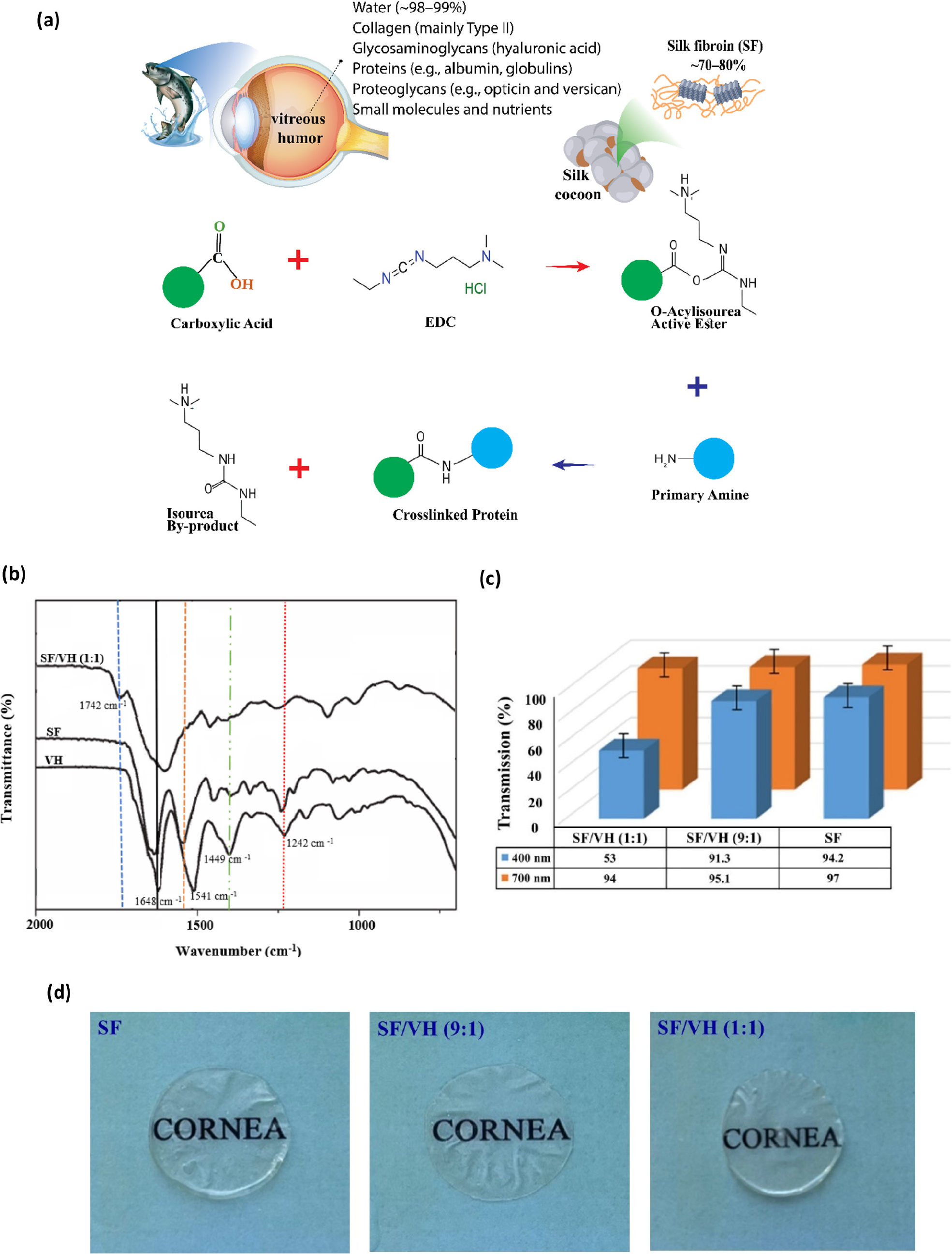
Fig. 1.
Schematic illustration of membrane preparation and EDC cross-linking (a), FTIR spectra (b), light transmittance (c), and visual transparency of hydrogels (d). VH: Non-cross-linked pure vitreous humor. SF, SF/VH (9:1) and SF/VH (1:1): EDC cross-linked hydrogel membranes made of pure silk fibroin, and SF/VH blends containing 90 % and 50 % silk fibroin, respectively.
FTIR analysis was conducted to examine the chemical bonds and functional groups present in the materials [Fig. 1(b)]. The freeze-dried VH, SF, and their blends revealed characteristic amide bands in all samples, confirming their proteinaceous nature. According to the spectra, the Amide I bands appeared at approximately 1,640 cm-1 in SF40) and VH,41) while the Amide III bands were prominent in VH and SF around 1,230-1,300 cm-1.41) Carboxylic groups related to GAGs, such as hyaluronic acid present in the VH are clearly visible in the 1,450 cm-1 band. Compared with the spectra of SF and VH samples, the peak at 1,742 cm-1, the characteristic band of ester, is distinctly increased in the spectra of the cross-linked sample, which indicates that cross-linking reactions might occur in the blend sample. Moreover, the spectra of SF and VH all show characteristic amide I and amide II peaks, but the cross-linked sample shows one peak at 1,605 cm-1, which means that the peaks of amide I and amide II overlap in the cross-linked hydrogels. Considering that the amide I band shifts to lower wave numbers during the formation of hydrogen bonds, while the amide II band shifts to higher wave numbers, the result indicates that many hydrogen bonds exist in SF and VH molecules.37) The intensity of the peak at 1,450 cm-1, related to carboxylic groups of hyaluronic acid in the VH was reduced in the cross-linked samples, which could be due to the reaction of the cross-linker with the carboxylic and amine groups and the formation of new amide groups. These compositional and conformational differences between fish-derived VH and SF, together with EDC cross-linking, are expected to significantly influence the formation of the hydrogel network and its functional properties, most notably optical transparency, which is essential for ocular applications.42)
For optimal visual acuity, the cornea must maintain both high light transmittance and transparency.43) The optical properties of the hydrogel membranes were analyzed using UV-Vis spectroscopy in the wavelength range of 400-700 nm. As expected, an increase in SF content resulted in enhanced light transmittance across the visible spectrum, particularly at higher wavelengths [Fig. 1(c)]. Similar behavior has been reported for collagen/SF composite membranes derived from fish skin and bovine tendon.16,43) Pure SF membranes also exhibited the highest transmittance, consistent with previous findings.43) Both pure SF and SF/VH (9:1) blend membranes showed excellent light transmittance exceeding 90 % across the visible range, comparable to that of the natural human cornea. The high transparency of the SF-VH hydrogels is likely due to the homogeneous distribution of components and the smooth, defect-free structure of the films, which reduces light scattering. Visual observations [Fig. 1(d)] further confirmed this trend, showing increased optical clarity with higher SF content. While molecular-level organization of protein chains may contribute to transparency, this study does not provide direct structural evidence for such alignment.
One of the key characteristics of polymer scaffolds is their ability to swell in biological environments, which facilitates the transport of nutrients and waste. The swelling degree of hydrogels highly depends on their cross-linking density. The swelling behavior of hydrogel scaffolds composed of SF and fish VH was evaluated in PBS at 2, 4, and 24 h [Fig. 2(a)]. All scaffolds exhibited a rapid uptake of the aqueous medium within the first 2 h, followed by a slower increase up to 24 h. The swelling ratio of the SF/VH blends was approximately 2-3 times higher than that of pure SF across all time intervals and increased proportionally with the VH content.
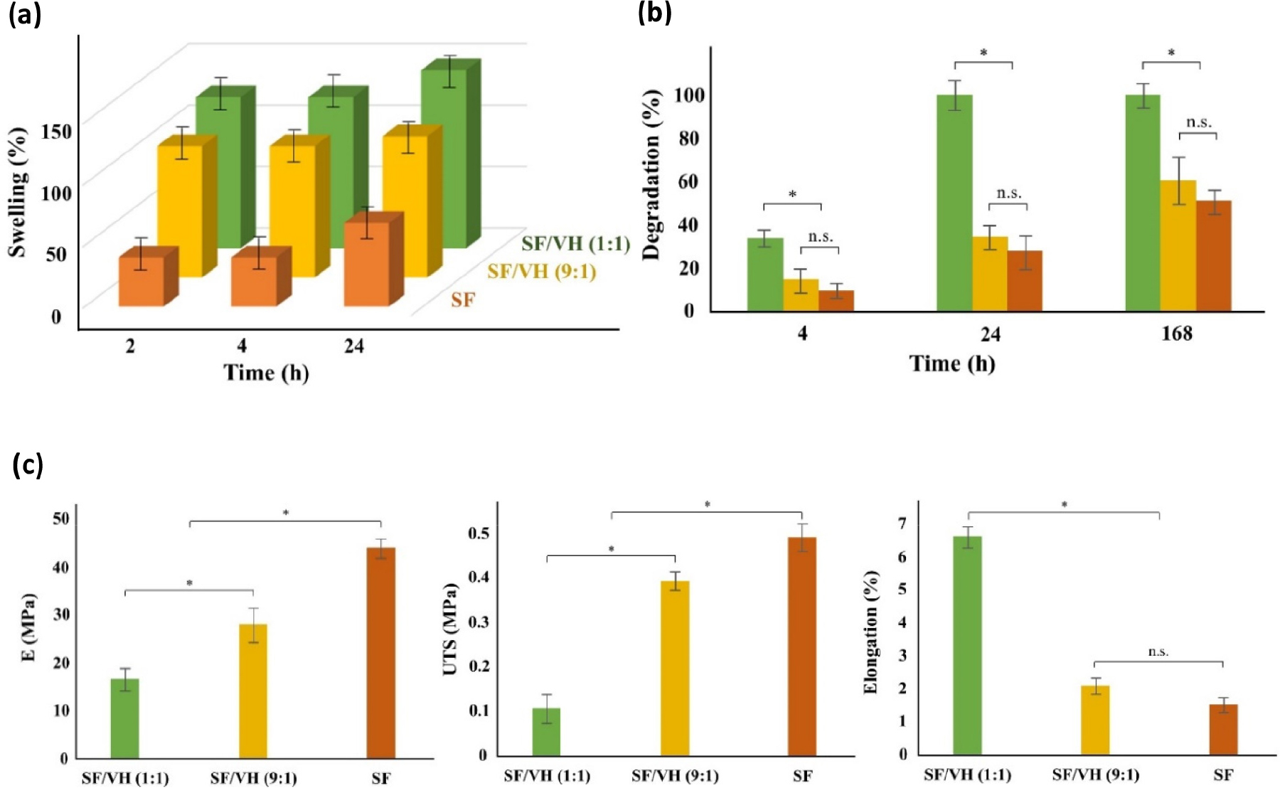
This positive correlation between water absorption and vitreous content can be attributed to differences in the cross-linking density of the samples. As previously described, EDC-mediated cross-linking occurs through reactions between carboxyl and amino groups of protein molecules, forming secondary amide bonds. Under a constant amount of cross-linker, the overall cross-linking density depends on the total protein content of the system. Since SF is highly proteinaceous compared to VH, an increase in SF content results in a higher cross-linking density and lower water uptake. Conversely, the lower protein content in VH leads to fewer available reactive sites for EDC, yielding networks with reduced cross-linking and enhanced swelling capacity. It is noteworthy that, under identical cross-linking conditions, pure VH could not form stable hydrogels or membranes, confirming its limited ability to undergo EDC-mediated cross-linking. After 24 h, the SF/VH (1:1) hydrogel showed the highest swelling ratio (~145 %), whereas the SF/VH (9:1) membrane also displayed a considerably higher swelling ratio compared to the pure SF membrane (117 % vs 70 %). Scaffold degradation is a critical factor in tissue engineering, as it should ideally match the rate of new tissue formation. Fig. 2(b) illustrates the enzymatic degradation of films with different SF/VH ratios when exposed to proteinase K over defined time intervals. The results demonstrated that SF was more resistant to enzymatic degradation than fish VH, and the overall degradation rate increased in proportion to the VH content. The proteinase enzyme requires access to the peptide bonds within the protein structure to break them down. SF contains numerous β-sheet structures, which are likely responsible for its high structural stability.42) These highly organized crystalline domains restrict enzymatic access to cleavage of peptide bonds, rendering them more resistant to proteolytic attack. In contrast, VH possesses a more accessible protein structure characterized by a high-water content, a gel-like network, weaker intermolecular interactions, and the absence of crystalline organization, which facilitates faster enzymatic degradation. Accordingly, the SF/VH blend scaffolds degraded more rapidly than pure SF, with degradation rates increasing with VH content. The SF/VH (9:1) hydrogel exhibited slower degradation compared to the 1:1 blend, indicating that higher SF content enhances structural stability. Notably, the 1:1 blend was completely degraded after one day, whereas the 9:1 blend retained approximately 40 % of its mass after one week, comparable to that of the pure SF sample, which showed about 52 % remaining mass. Mechanical testing further confirmed the compositional effects on the membrane properties, demonstrating that increasing the vitreous content reduced the mechanical strength while improving flexibility. SF is a natural structural protein that forms the core of silk fibers, renowned for its high strength and toughness. Its mechanical robustness originates from repetitive glycine-alanine-glycine-alanine-glycine-serine (GAGAGS) sequences that self-assemble into antiparallel β-sheet structures. These highly ordered crystalline domains cross-link the protein chains through extensive hydrogen bonding and van der Waals interactions, conferring exceptional tensile strength and rigidity to the material.44) In contrast, fish VH is a semi-liquid, gel-like material composed of approximately 99 % water, with the remainder consisting mainly of hyaluronic acid and proteins.45)
When blended, VH acts as a plasticizer, softening the SF matrix by intercalating between the amino acid chains of the SF protein, thereby reducing intermolecular forces and increasing chain mobility. As shown in Fig. 2(c), incorporation of 10% VH decreased the Young’s modulus from 43.9 MPa in pure SF to 27.9 MPa in the SF/VH (9:1) blend, while the ultimate tensile strength (UTS) decreased from 0.49 MPa to 0.39 MPa. Further increasing VH content to 50 wt% (1:1 blend) led to a pronounced loss of mechanical strength, resulting in mechanically weak membranes. Conversely, the strain at break increased with higher VH content, from 1.5 % in pure SF to 2.1 % in the 9:1 blend, indicating improved flexibility.
For comparison, the native human cornea exhibits a Young’s modulus of approximately 0.3-3 MPa (Table 1) (hydrated anterior stroma) and a UTS of 0.3-1 MPa, with elongation at break around 6-10 %. Although the SF/VH (9:1) blend is stiffer than the native cornea, it maintains a balance of mechanical strength and elasticity, providing sufficient support while allowing some flexibility. In contrast, higher VH content (50 wt%) reduces stiffness and strength too much, potentially compromising structural integrity. These results indicate that moderate VH incorporation (10 %) offers an optimal compromise between mechanical reinforcement and flexibility, aligning the hydrogel properties closer to the physiological requirements of corneal tissue.
Table 1.
Main mechanical properties of the native human cornea.
Biocompatibility of the hydrogel scaffolds was evaluated using the MTT assay, which demonstrated excellent cellular responses [Fig. 3(a)]. The viability of HCECs cultured on pure SF, SF/VH (1:1), and SF/VH (9:1) blend hydrogels, as well as pure fish vitreous samples, was assessed after 24, 48, and 72 h. Cell adhesion was examined at 24 h as an early stage of cell growth, while cell proliferation and colonization within the hydrogels were evaluated at 72 h. All samples exhibited non-cytotoxic behavior throughout the culture period, with cell proliferation increasing over time. Pure SF hydrogels showed good biocompatibility (80-90 % viability) with no significant difference compared to the TCPS control. Incorporation of VH enhanced cell viability in a concentration-dependent manner, with the addition of only 10 % VH increasing viability to approximately 115 %. Higher VH contents further promoted cell growth, with the number of viable HCECs nearly doubling compared to the control at all time points. The pure VH samples showed the highest proliferation, reaching ~220 % after 72 h, likely due to the presence of various growth factors that stimulate cell proliferation. The vitreous fluid serves as a reservoir for numerous locally produced ocular growth factors. These include transforming growth factor-beta (TGF-β), fibroblast growth factors (FGFs), insulin-like growth factors (IGFs), epidermal growth factor (EGF), and vascular endothelial growth factor (VEGF). These biomolecules act through autocrine and paracrine mechanisms to support the maintenance, survival, and differentiation of ocular tissues, particularly contributing to retinal vascularization and neurogenesis.46)
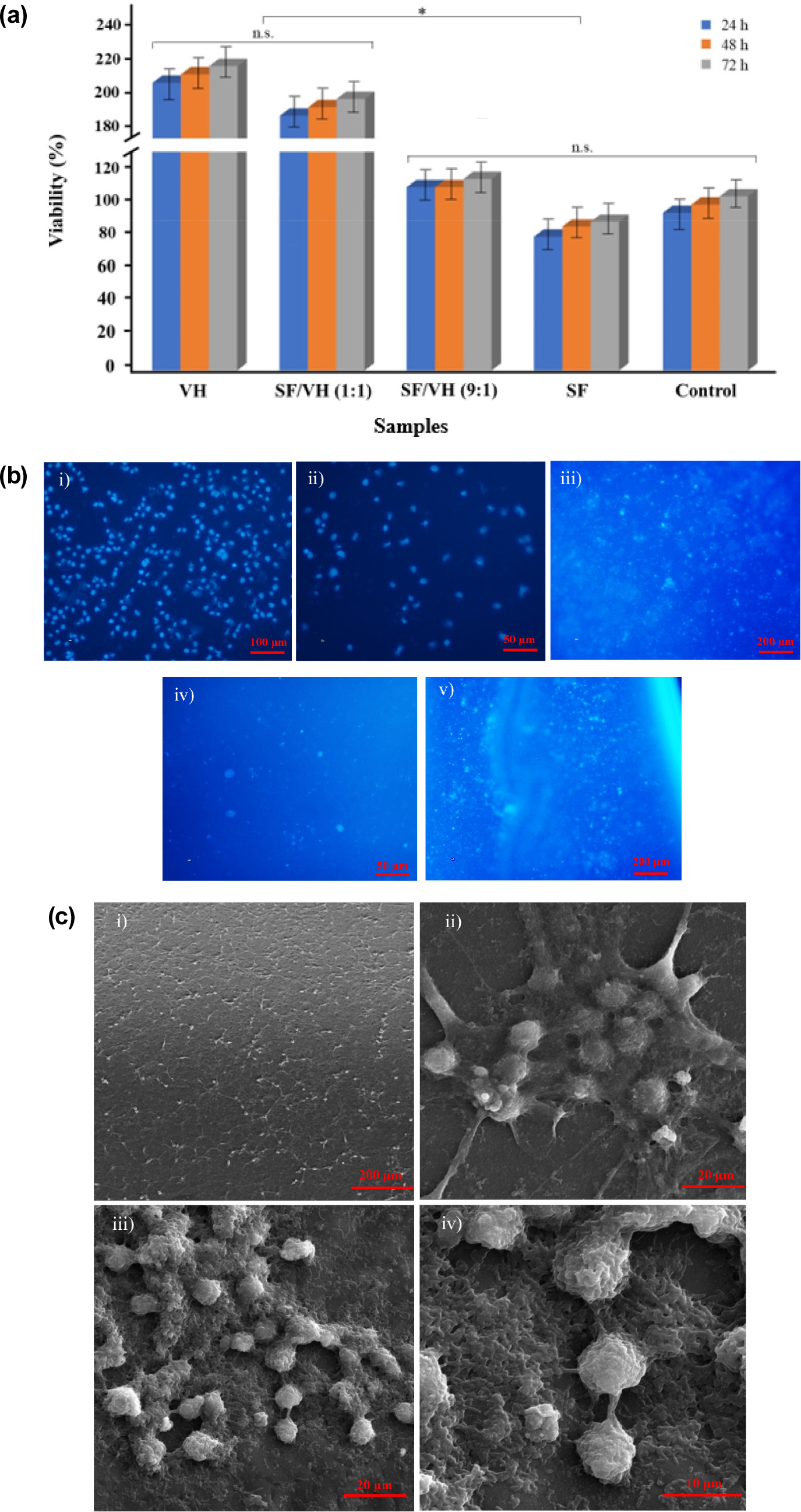
Fig. 3.
(a) 24-72 h viability of HCECs; (b) DAPI images of HCECs cultured with the hydrogel samples: (i) VH, (ii) SF/VH (1:1), (iii) SF/VH (9:1), (iv) SF; (v) control (TCPS); (c) SEM images illustrating HCECs attachment on the surface of SF sample (i and ii) and SF/VH (9:1) (iii and iv) at different magnifications. *p < 0.01, n.s.: not significant.
Consistent with the MTT results, DAPI staining [Fig. 3(b)] revealed a higher number of nuclei in VH-containing samples, confirming enhanced proliferation proportional to VH concentration. Despite the superior proliferation observed in VH-rich formulations (i.e., pure VH and the 1:1 blend), only 10 % VH was sufficient to produce a pronounced positive effect in both MTT and DAPI analyses. Therefore, the SF/VH (9:1) hydrogel demonstrated an optimal balance between favorable biological performance and material stability. SEM images of the SF/VH (9:1) hydrogel further confirmed excellent cell adhesion and spreading, with distinct cytoplasmic extensions and greater cellular aggregation compared to the pure SF hydrogel substrate [Fig. 3(c)]. Given the excellent results of the cell studies, as bioengineered constructs derived from animal sources may still provoke allergic or immunogenic responses. Clinical investigations are essential to assess long-term safety, biocompatibility, and potential immune effects before clinical translation.
4. Conclusion
In this study, SF and fish VH were combined and cross- linked using EDC to fabricate transparent, biocompatible hydrogel membranes for corneal epithelial tissue regeneration. Comprehensive physicochemical, mechanical, and biological evaluations demonstrated that the blend composition has a significant influence on scaffold performance. Increasing VH content enhanced flexibility, swelling, and cell proliferation, whereas higher SF content improved transparency, stability, and mechanical strength. Both pure SF and SF/VH (9:1) membranes exhibited excellent light transmittance (> 90 %), comparable to the natural human cornea. Cytocompatibility studies confirmed non-toxicity for all formulations, with the SF/VH (9:1) hydrogel providing an optimal balance of transparency, mechanical performance, and biological activity. These findings suggest that incorporating small amounts of VH into SF matrices can enhance bio-functionality while maintaining structural and optical properties. However, as this study is limited to early-stage in vitro evaluation, definitive conclusions regarding clinical applicability cannot yet be drawn.
Future studies should investigate long-term in vitro stability, in vivo performance, immune compatibility, and potential allergic reactions. Additionally, scaling up production, exploring alternative marine VH sources, and integrating growth factors or bioactive molecules could further improve the utility of SF/VH hydrogels for corneal tissue engineering.
