


1. Introduction
The scintillator is essentially a luminescence material that absorbs the high energy photons and then emits visible light. The cerium ion was studied as a fast luminescence center in many hosts. The cerium doped silicate based scintillators, such as LSO(Lu2SiO5),1) LPS (Lu2Si2O7),2) LYSO(Lu2Y2SiO5),3) and GSO(Gd2SiO5),4) have been developed. These material are used for gammaray detection in positron emission tomography(PET), astrophysics, nuclear medical diagnostic instruments, and high energy and nuclear physical experiments. Therefore, the investigation on the growth and scintillation properties of ceramic scintillator materials has become an attractive and interesting field in the past decade. Lutetium based crystals are grown by Czochralski method,3,5-7) of which the cost is very high, and the maximum reachable concentration of rare earth ion is quite low. The advantages of the sol-gel process are the possibility to prepare easily thin films (or powder) from solution and it is very attractive.8-12)
Lutetium-based materials have been of concern for the last few years, in which cerium(Ce) doped of Lu2SiO5(LSO),1,9,12) Lu3Al5O12(LuAG),13) LuBO314) and LYSO15) have become the most competitive candidates to replace Bi4Ge3O12(BGO) in positron emission tomography(PET). Due to its high density of lutetium oxide, lutetium-based materials have been considered as a promising host matrix of rare earth ions to produce phosphors and scintillators. In particular, cerium doped lutetium silicate(LSO and LPS) are the most stable compounds, and seem to be very efficient scintillator. However, the ion radius of cerium (1.034 Å) is much larger than that of lutetium(0.86 Å), it is expected to be difficult to incoporate a large amount of cerium into the lattice of lutetium based material. Yan et. al suggested that the solubility of serium ion in the LPS host lattice is higher than in the LSO lattice.5) We have been preparing LSO:Ce3+ and LPS:Ce3+ powders through a sol-gel process, properties of sol-gel derived LSO:Ce3+ powders have been reported recently.12) However, Lu2Si2O7: Ce3+ materials have been synthesized rarely using sol-gel method, and the scientillator characteristics are not fully understood.
In this paper, Lu2Si2O7:Ce3+ powders were synthesized by the sol-gel method. The morphology, crystal structure, photoluminescence characteristics of sol-gel derived Lu2Si2O7:Ce3+ powders were investigated.
2. Experimental Procedure
Cerium doped Lu2Si2O7 powder was synthesized by sol-gel process at room temperature. Lutetium nitrate hydrate( Lu(NO3)3·nH2O), tetraethyl orthosilicate(Si(OC2H5)4), cerium nitrate hydrate(Ce(NO3)2·xH2O) and i-C3H7OH (Showa chemical Co. Ltd., Japan) were used as the starting materials. At first, Si(OC2H5)4(TEOS) was hydrolyzed, vigorously stirring at 40 °C for 1hr after dissolving it into the 10 ml of i-C3H7OH solution. Lutetium nitrate hydrate(Lu(NO3)3·6H2O) and cerium nitrate hydrate(Ce (NO3)2·xH2O) were dissolved separately in 10 ml of i- C3H7OH, and then two solutions were mixed together. The mixed solution was vigorously stirred at 25 °C for 1hr, and a clear precursor sol was obtained by completely mixing the reactants. The transparent solution was allowed to form a gel at 60 °C and the gel was dried at 110 °C for 24h. A gel was formed after a few days, of which color was turned into light yellow and transparent. This gel was then calcined at 1,200 °C for 2h in order to obtain Lu2Si2O7:Ce3+ crystalline powder. During heating, the temperature was held at 300 °C for 2h to remove the residual organic compounds. The preparation process of the Lu2Si2O7:Ce3+ powders is shown in Fig. 1 and were prepared according to our previous work.12)
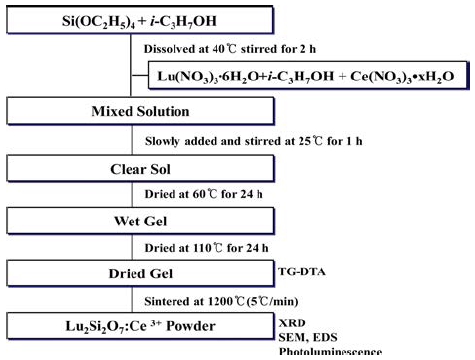
The crystal structure of the powder was examined, using X-ray diffractometer(XRD, D/Max-220, Rigaku, Japan) with Cu Ka radiation operated at 40 kV and 30 mA. The thermal behavior of the dried gel was investigated, using TG-DTA(STA 409, Netzsch, Germany), and carried out at up to 1,200 °C in air at a constant heating rate of 5°/ min, using Al2O3 as a reference material. The surface morphology was observed, using scanning electron microscope (SEM, Hitachi, S-4300, Japan) and EDS(Horiba EMAX 2770). Emission spectra were measured, using fluorescence spectrophotometer(Perkin-Elmer, LS-55B, USA).
3. Results and Discussion
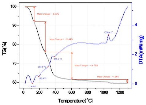
The crystallization of cerium doped Lu2Si2O7 powder was studied by thermal analysis, which is shown in Fig. 2. A sharp exothermic peak is observed at 1,051 °C and corresponds to the crystallization of Lu2Si2O7 phase. It shows that the phase transformation in the cerium doped Lu2Si2O7 phase is lower than in the cerium doped Lu2SiO5 phase (at 1,073 °C).12) Because the Lu2Si2O7 phase shows a more open lattice in the Lu2SiO5 phase,5) the phase transition is a similar to cerium doped Lu2SiO5 powder. Total weight loss is approximately 40 %. It is important to note that this crystallization temperature is well below temperatures required by the Czochralski method (about 1900 °C) and in comparing with the solid state reaction temperature (about 1400 °C). The cost of starting materials is lower and the melting point is lower in LPS crystal (about 1900 °C) than in LSO crystal (about 2100 °C ).5)
The cerium doped Lu2Si2O7 powders were calcined at an interval of 200 °C in range of 800 °C to 1,200 °C and the crystallization phase composition was identified with XRD, which is shown in Fig. 3. The gel powder remained amorphous up to 800 °C. It was seen that Lu2O3 phase appeared at 800 °C. The crystallinity was increased as the heating temperature was increased. At 1,000 °C the all characteristics peaks of Lu2Si2O7 phase appeared, and any other phases were not formed. When the calcination temperature reaches 1,200 °C, continued refinement of peak shapes and intensities were observed, indicating crystallite growth of the Lu2Si2O7 phase with the calcined temperature increasing. The formation of Lu2Si2O7 phase was confirmed by XRD analysis for the powder heated at 1,200 °C. The Lu2Si2O7 powder is in agreement with the Lu2Si2O7 crystal reported(JCPDS card No. 35-0326).(5,6)
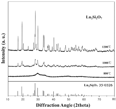
Fig. 3
X-ray diffraction patterns of the Lu2Si2O7:Ce3+ powder calcined at various temperatures for 2h.
Fig. 4 shows the morphology and EDS of cerium doped Lu2Si2O7 powders which were synthesized by solgel process and calcined at 1,200 °C for 2h. Cerium doped Lu2Si2O7 powders are slightly agglomerated because of crystallite growth at high calcined temperature. Cerium doped lutetium pyrosilicate(LPS) powder was homogeneous and constituted of small spherical particles of about 500 nm. Fig. 4(right) shows that the surface of gel powders was doped efficiently with cerumion.
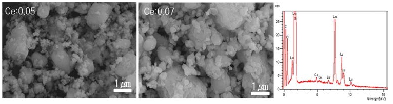
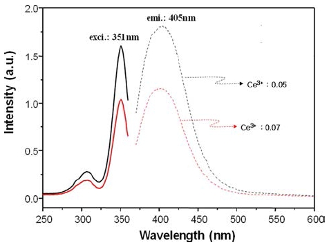
Fig. 5 shows the excitation and emission spectrum of Lu2Si2O7:Ce3+ powders with the cerium ion contents (0.05 mol% and 0.07 mol%). It is clear that the excitation spectrum is essentially identical to the absorption spectrum. The excitation spectrum of Lu2Si2O7: Ce3+ powders taken at emission of 405 nm show two bands which have peaks at 306 and 351 nm, which are assigned to the Ce3+ ion transiting from the 5d level to the 4f ground states. The emission spectrum of Lu2Si2O7:Ce3+ powders excited at 351 nm are shown in Fig. 5. The emission intensity increases at a cerium doping of 0.05 mol%, but decreases at 0.07 mol%. The emission spectrum shows a broad band in range of 350-525 nm, and the broad wavelength band on the right side of the spectra should be ascribed to the same 5d-4f transitions of Ce3+ as the case of Ce doped Lu2Si2O7:Ce3+ single crystal.5,7)
4. Conclusion
Cerium doped lutetium pyrosilicate(LPS) powders were prepared by sol-gel process with different cerium contents (0.05 mol% and 0.07 mol%). The formation of lutetium pyrosilicate(LPS) phase was confirmed by XRD analysis for the powders heated at 1,200 °C, in which single phases of Lu2Si2O7 was observed. Lutetium pyrosilicate(LPS) powder was homogeneous and constituted of small spherical particles of about 300 nm. The excitation spectrum of Lu2Si2O7:Ce3+ powders taken at emission of 405 nm shows two bands peaking at 306 and 351 nm, which are assigned to the Ce3+ ion transiting from the 5d level to the 4f ground states. The emission intensity increases at a cerium doping of 0.05 mol%, but decreases at 0.07 mol%. The emission spectrum shows a broad band in range of 350-525 nm, and the broad wavelength band on the right side of the spectra should be ascribed to the same 5d-4f transitions of Ce3+ as the case of Ce doped Lu2Si2O7:Ce3+ single crystal.